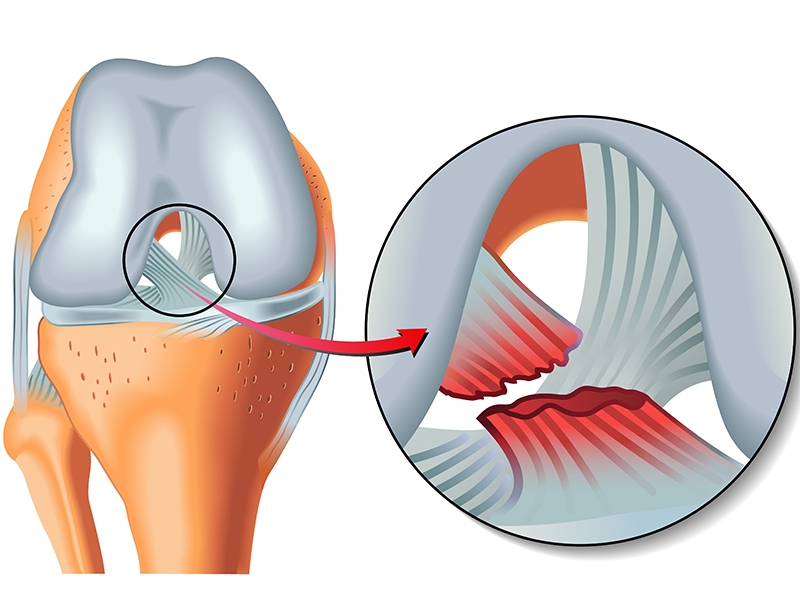
Anterior Cruciate Ligament Rupture: Anterior Cruciate Ligament (ACL) is one of the stabilizators of the knee joint. It’s main function is preventing excessive forward tibial sliding under the femur. ACL rupture is a relatively common injury of the knee joint. It mostly occurs in sport injuries. The mechanism of injury is usually a non contact rotational forcing of the knee joint. Patient usually hears or feels a pop coming from the knee joint. Although usually not intense, pain is common. Knee swelling is progressive. A knee joint swelling within 24 hours after a rotation injury is indicative of ACL rupture in 80% of the cases. Progression of the swelling increases pain and causes knee joint motion limitation. Besides ACL rupture, there may be associated lesions like meniscus tear or cartilage lesions. Initial treatment includes knee immobilization with brace, elevation, compression and cold application. Physical examination at the early phase may be difficult due to swelling and pain. MR examination gives the best diagnosis.
ACL rupture doesn’t heal with medication, immobilization or physical therapy. Surgical reconstruction of the Ligament is the only treatment in required cases.
To whom the surgical reconstruction should be done? Surgery should be done in all active, sporting young individuals. ACL deflcient knee may cause associated injuries to knee joint and early cartilage erosion. An individual who wants to continue active sporting life must have reconstruction of the ACL. Another indication for ACL reconstruction is feeling sliding of the knee joint during daily activities like climbing stairs or knee bending. This indicates that there is an abnormal motion of the knee joint even with minor activities.
When to reconstruct the ACL: It is more beneficial to wait for regression of the joint effusion and to gain full knee range of motion. This usually takes 1 month after the injury. During this time patient is advised to begin physical therapy to gain muscle strength which will be very useful after the surgery.
How is the surgery?
Surgery is done arthroscopically without opening the joint. Two hamstring tendons located on the medial side of the joint is removed and a quadruple loop is made. Two tunnels are made, one through the tibia, the other through the femur. The hamstring tendons are passed through the tunnels and fixed with special endo-buttons and absorbable screws and staples. Surgery usually takes 1,5-2 hours. Associated injuries like meniscus tears are fixed at the same stage.
What happens after the operation?
Patient is usually hospitalized for 1 night for pain control or sometimes discharged at the post- operative day. A brace is usually advised for 3-4 weeks for immobilization and gradual weight bearing with crutches is allowed. Physical therapy begins 3-4 days after the surgery to regain muscle strength and knee joint motion. Therapy continues 3-4 weeks than after the patient is advised to continue muscle strengthening exercises. Active high level sport actlvlties are restricted for 6 months.